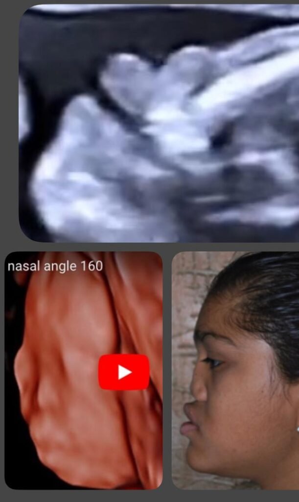
Binder syndrome fetal ultrasound by Dr.Noor Beskri
Binder syndrome, also known as nasomaxillary dysplasia or maxillo-nasal dysostosis.
Although the true etiology is unknown, it is most likely multifactorial,The current understanding is that Binder syndrome is not a disease but rather a phenotype with several causes, of which the most important is chondrodysplasia punctata.
Binder phenotype is associated with either :
•chondrodysplasia punctata(Binder syndrome is the mildest form of chondrodysplasia punctata(isolated facial characteristics ).
•Keutel and Nager syndrome.
•Maternal :intake of anticoagulants(especially warfarin),systemic lupus,*hyperemesis gravidarum and environmental circumstances affecting vitamin K metabolism.
•36 percent of the individuals had a positive family history,This points to an autosomal recessive inheritance pattern with little penetrance or multifactorial inheritance.
Binder’s syndrome affects both men and women equally.
There have been rare instances of family recurrence of the syndrome (among siblings or parents and children).
It has distinct impacts on facial characteristics. A “dish-face” deformity and a retruded flat nose describe this congenital abnormality but it supports the idea that this rare facial syndrome develops in the context of skeletal dysplasia.
Binder syndrome is characterized by:
Fetal facial dysmorphism ,arhinoid
face, absence of frontal sinus + abnormal hypoplastic nasal bones,deviated septum,Absent anterior nasal spine,maxillary hypoplasia .a shortened nose with depressed nasal bridge, flat facies and a convex upper lip. Malocclusion, underdevelopment of frontal sinus and anomalies of the cervical spine may be present . Most of the individuals with Binder phenotype have skeletal dysplasia, but the range of manifestations is wide, from mild to severe. While in some cases only the facial features are present, other cases display the full clinical picture of the X-linked brachytelephalangic chondrodysplasia punctata.
Diagnosis and Treatment of Binder Syndrome:
Similar facial features in other syndromes, such as Warfarin embryopathy, Down syndrome, Apert syndrome, and others, are used to make the differential diagnosis for Binder’s syndrome.
During fetal ultrasonogram (USG) exams, these syndromes are frequently misdiagnosed. Beginning in the 21st week of pregnancy, Binder’s syndrome can be easily detected with 2D and 3D ultrasounds. Nasal abnormalities are found in 1 in 1600 fetuses and may play a significant role in the early detection of congenital illnesses and syndromes.
It’s also worth noting that Down syndrome is frequently linked to Binder’s syndrome. According to reports, around 12% of patients with trisomy 21 do not have a nasal bone. The score is much higher among aborted Down syndrome fetuses, with abnormalities in the nasal bone found in 60% of those (in 26%, the nasal bone is absent, and in 36%, it is hypoplastic).
*Treatment planning necessitates nose reconstruction first and foremost, although additional medical services (such as orthodontic and orthognathic treatment) may also be required. The child should be placed under the care of a plastic surgeon and an orthodontist right after birth. The timing and types of surgeries used to treat nasomaxillary dysplasia patients are determined by the degree of the deformity and are scheduled individually.
References:
- Hunt JA, Hobar PC. Common craniofacial anomalies: the facial dysostoses.
2 .Keppler-Noreuil KM, Wenzel TJ. Binder phenotype: associated findings and etiologic mechanisms
And others.


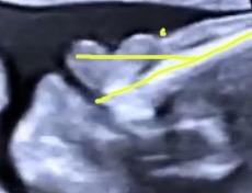
Ultrasound presentation of Binder syndrome – sagittal view showing the nasal and frontal bones (yellow lines), flat face with fronto-nasal angle (•) above 130°

