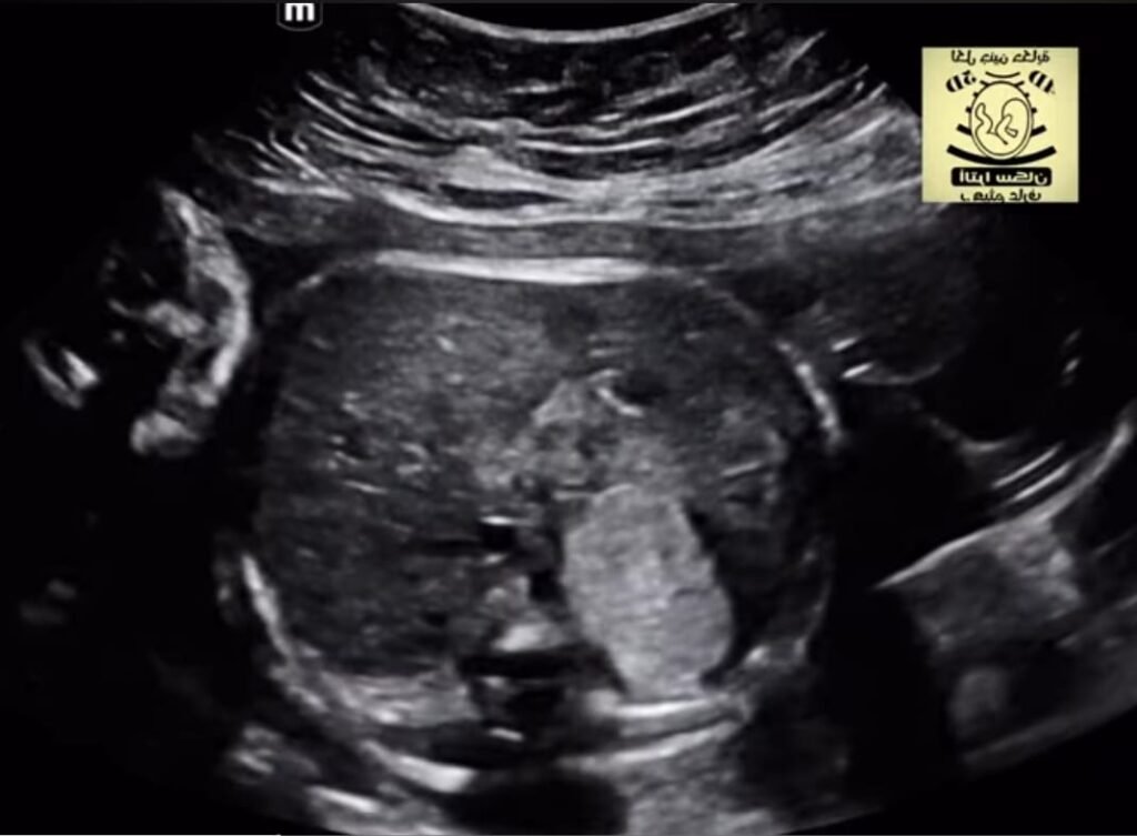
Extralobar pulmonary sequestration
More common in males (male-to-female ratio, 4:1), an extralobar sequestration can be seen on fetal sonography as early as 16 weeks’ gestation and typically appears as a solid well-defined triangular echogenic mass
The extralobar sequestration may be supradiaphragmatic (90%) or subdiaphragmatic (10%).
Most supradiaphragmatic sequestrations occur between the left lower lobe and diaphragm; most subdiaphragmatic sequestrations are also left-sided.
Cysts can be seen, particularly in hybrid lesions that combine elements of sequestration and congenital cystic adenomatoid malformation
Visualization of a systemic feeding artery
arising from the thoracic or abdominal aorta is a useful finding that distinguishes a sequestration from other masses such as a congenital cystic adenomatoid malformation or bronchial atresia. Color and spectral Doppler sonography can be helpful in visualizing the feeding artery, but visualization may still be difficult. In one study, a feeding systemic artery was identified on Doppler sonography in only four of 10 cases of pathologically proven sequestration .