Interrupted Aortic Arch (IAA) FETAL ECHO
🫀 THE WHOLE ARTICLE IS BY MY DEAR FRIEND DR COSKUN UMIT MFM SPECIALIST FROM TURKEY
i reached the diagnosis thanks to him
Interrupted Aortic Arch (IAA) is a rare but critical congenital heart defect characterized by a
discontinuity between the ascending and descending aorta. Prenatal diagnosis is essential for optimal perinatal planning and improved outcomes.
🫀 The three-vessel and trachea (3VT) view plays a crucial role in the fetal echocardiographic evaluation of IAA. This axial plane provides a comprehensive overview of the great vessels in relation to the trachea. In a normal 3VT view, the aortic arch and ductus arteriosus appear as two continuous, forward-flowing vessels forming a V-shaped confluence to the left of the trachea.
🫀 In cases of IAA, this pattern is disrupted. Sonographic clues include:
● Absence or discontinuity of the aortic arch in the 3VT view (V shape can not be formed)
● A prominent and isolated ductus arteriosus
● Very small aorta (smaller than SVC)
● A right-sided or midline trachea in relation to the ductus
🫀 Recognition of these subtle but significant changes in the 3VT view can raise early suspicion for IAA and prompt detailed evaluation of the aortic arch anatomy and associated intracardiac anomalies.
🫀 Early and accurate detection of IAA on fetal echocardiography is key to ensuring appropriate neonatal management, including timely surgical intervention.
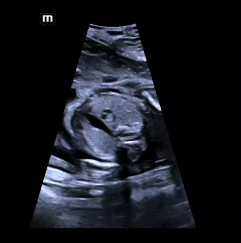
🫀 Here is a 23 weeks poor fetus with Interrupted Aortic Arch (note the very small aorta).