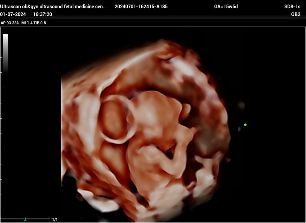
occipital encephalocele fetal ultrasound by Dr.Sara Salem, Lybia
مقاله بقلم الدكتوره ساره سالم من ليبيا
*
Occipital Encephalocele
The herniated structure
looks consist of meninges only (meningocele) e’out cerebral tissue.* • A variant of neural tube defects, involve herniation of intracranial structure w’ occur as a result of skull defect due to Failure of closure of the cranial end of the ectodermal neurotube.

• it occur very early in embryogenesis As skull
ossification starts at 10 weeks’ gestation.• _Once an encephalocoele is diagnosed:_ ✔️ Search for other anatomic abnormalities (40% had associated congenital anomalies mostly extra-CNS malformations) • The spine should be examined to exclude associated spina bifida.• The fetal kidneys should be examined, because of a high incidence of association with renal cystic disease (Meckel–Gruber syndrome). _favorable outcome seen e’:_ ✔️ meningeal herniation only✔️ presence of normal karyotype ✔️ lack of associated CNS or other malformations (i:e isolated encephalocele).• _Post natal_ : Surgical repair of encephalocele, generally timed for 0 to 4 months of age.https://youtu.be/y2df4RreIv4?si=wYXc5OJzS3RwujZB

