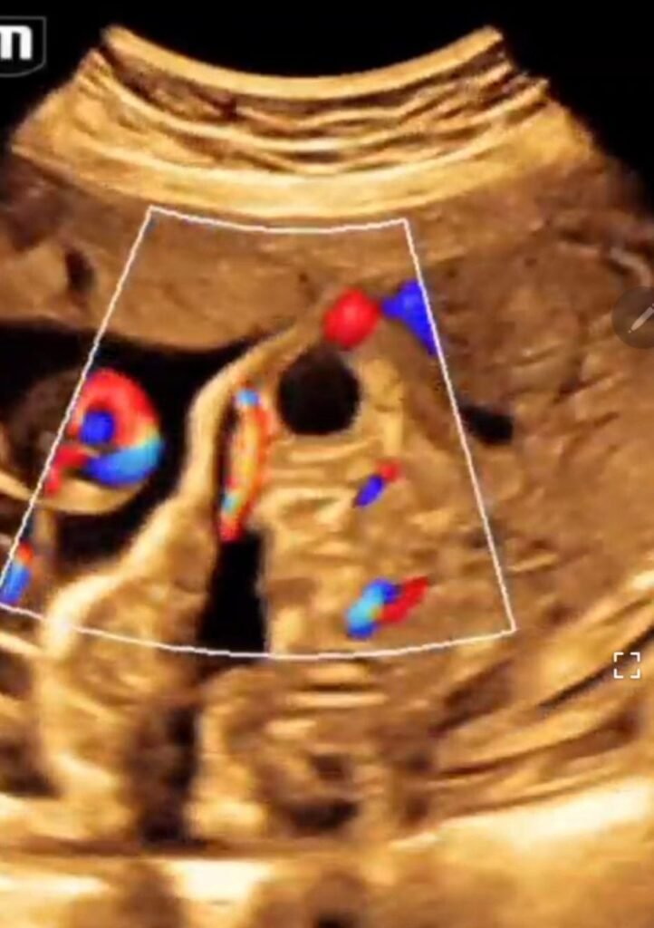
URACHAL CYST FETAL ULTRASOUND by Dr.Amira Shawky
🔘Urachus channel closes at both bladder & umbilicus but leaves a cyst ( fluid filled sac ) in the middle portion of the channel..
Urachal cyst is the most common type of Urachal anomalies …
🔘Persistent Urachus is a rare congenital anomaly. The urachus is a fibrous remnant of the allantois, connecting fetal urinary bladder (UB) to the umbilical cord (UC) and runs within the UC. The urachal cyst is a collection remaining due to lack of apposition of Allantoic sheets.
🔘fetus presented with anechoic cyst at the base of umbilical cord.
🔘A fetal urachal cyst refers to a urachal cyst occuring in utero. It may or may not communicate with the vertex of the fetal bladder. It may also arise within the umbilical cord. Umbilical cord urachal cysts originate from an extra-abdominal urachal system.
🔘rare anomaly with an estimated incidence of 0.25:10,000 deliveries1. Males are affected twice as commonly as females.
🔘The differential diagnosis of patent urachus includes anterior abdominal wall defects, bladder exstrophy, vascular lesions of umbilical cord (hemangioma, varix, true knot) or allantoic or omphalomesenteric cysts.

🔘Postnatal surgical resection and histopathology evidence of transitional epithelial lining confirmed the diagnosis of persistent Urachal cyst in either case. Early detection allowed for appropriate counselling and prompt corrective surgery after birth.
🔘Conclusion
The permeable urachus is a progressive disease that can be diagnosed by prenatal ultrasound as early as the first trimester. This condition does not justify additional imaging or karyotype studying. Surgical treatment is often necessary, but spontaneous resolution is possible during the pregnancy or after birth when the cysts are simple simple.

