Placental chorioangioma
beautiful collage by Dr ALAA OBEID
Placental chorioangiomas are benign vascular tumors of placental origin. It is the most common tumor of the placenta and is usually found incidentally.
Epidemiology
The estimated incidence is at ~1% of all pregnancies .
Associations
Recognized associations include:
- hydrops fetalis: also listed under complications
- polyhydramnios: also listed under complications
- Beckwith-Wiedemann syndrome
- single umbilical artery
- fetal anemia
- fetal congestive cardiac failure
- hepatic and cutaneous hemangiomas
- IUGR
Clinical presentation
In most cases, chorioangiomas are asymptomatic, and merely incidental findings. Occasionally, when they are large or multiple, they can result in poor outcomes for both the fetus and the mother.
Pathology
A chorioangioma is thought to arise as a malformation of the primitive angioblastic tissue of the placenta. The angiomas are perfused by fetal circulation and thus, when they are large, they may represent a significant impediment to fetal cardiac activity. They may also sequester platelets and can, in turn, give fetal thrombocytopenia.
There is some debate as to the exact nature of chorioangiomas. Most authors consider them as benign neoplasms, while others categorize them as hamartomas, given their composition of mostly native placental tissue and their inability to metastasize.
There can be significant variation in size. Most lesions tend to be small and lesions >4 cm are rare . Large tumors can, however, produce degenerative phenomena like necrosis, calcification, hyalinisation, or myxomatous degeneration.
Subtypes
Three histological types are recognized:
- angiomatoid: characterized by numerous blood vessels
- cellular: with poor vascularization
- degenerative
Location
They tend to occur on the fetal side of the placenta (close to cord insertion).
Markers
- raised maternal alpha-fetoprotein (MSAFP)
Genetics
Most cases tend to be sporadic.
Radiographic features
Ultrasound
Typically a chorioangioma is located near the insertion of the cord and protrudes into the amniotic cavity.
- often seen as a hypoechoic, rounded mass, located near the chorionic plate +/- umbilical cord insertion site
- usually contains anechoic “cystic” areas and can be seen as distinctly separate from normal surrounding placental tissue
- some heterogeneous areas caused by degenerative processes and internal hemorrhage can be seen
- can also rarely appear pedunculated
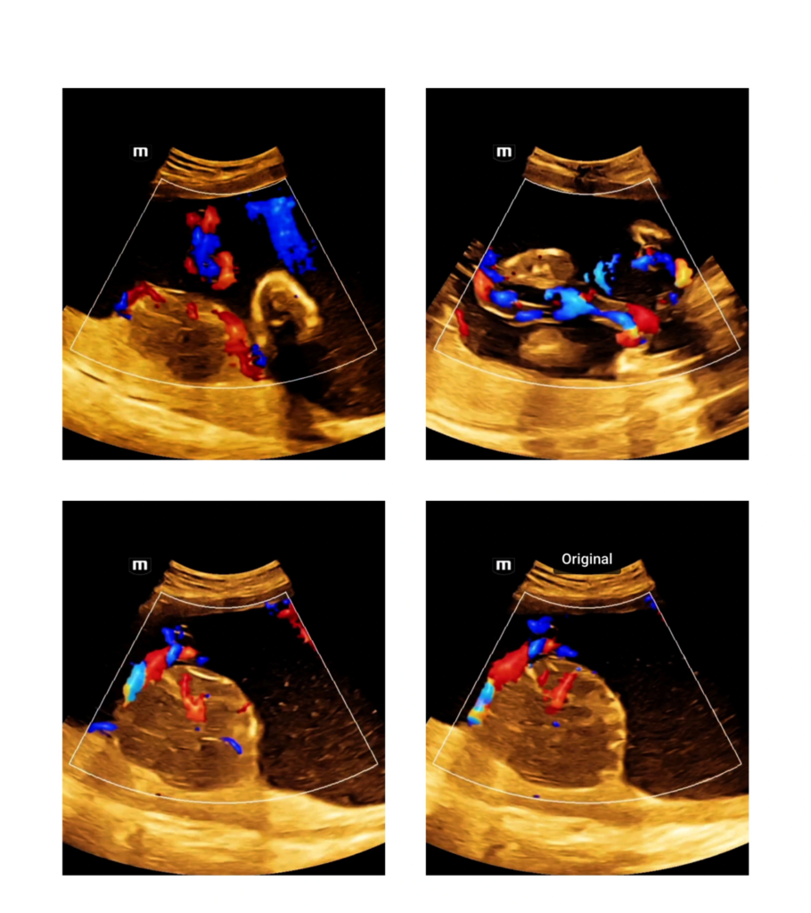
- on color Doppler, often demonstrates low resistance pulsatile flow within the anechoic “cystic” areas, which represent enlarged vascular channels
Large chorioangiomas may undergo spontaneous infarction with decreased echogenicity, decreased tumor volume, and decreased blood flow on color Doppler images .