
omphalocele fetal ultrasound
Omphalocele fetal ultrasound
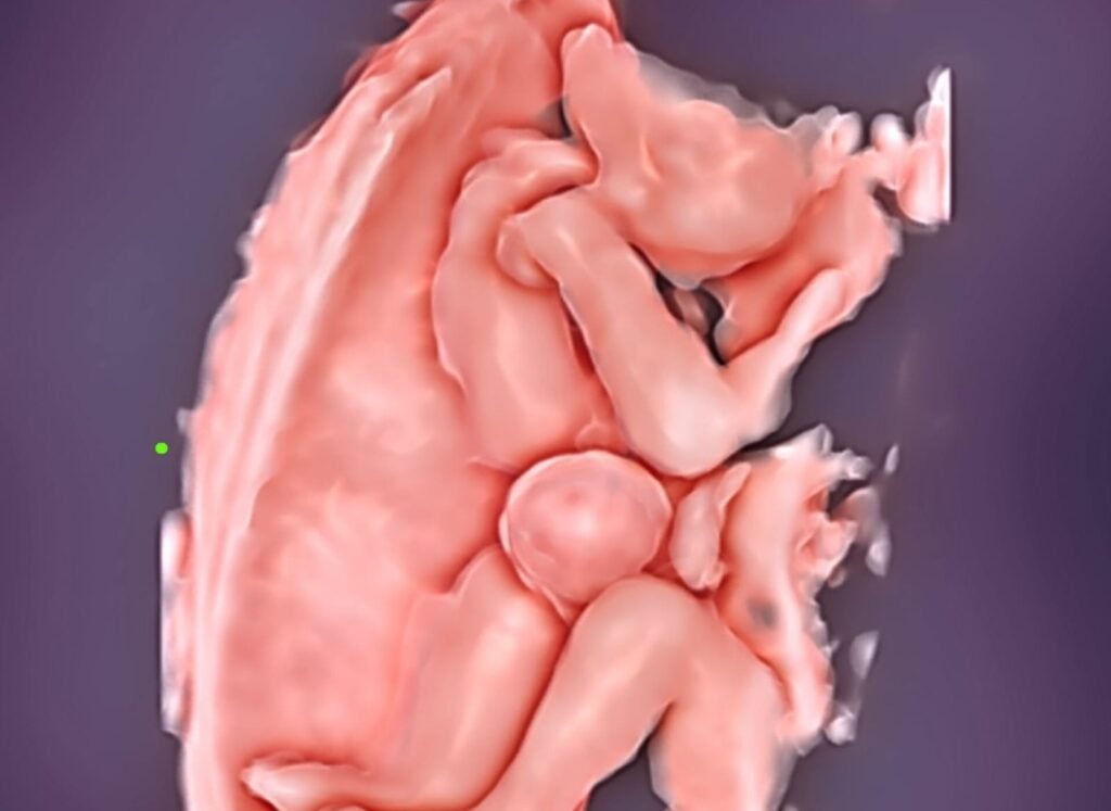
At 8-10 weeks all fetuses demonstrate herniation of the midgut that is visualized as a hyperechogenic mass in the base of the umbilical cord.
Retraction into the abdomen occurs at 10–12 weeks.
For an exomphalos containing bowel only, the prevalence is about 1:100 for crown-rump length (CRL) of 45.0-54.9 mm, 1:800 for a CRL of 55-64.9 mm and 1:2,000 for CRL of 65.084.0 mm.
The prevalence for an exom/phalos
containing liver is about 1:3,500.
Exomphalos containing liver persists throughout pregnancy, whereas exomphalos containing bowel only resolves by 20 weeks’ gestation in about 90% of cases.
Associated chromosomal
abnormalities, mainly trisomy 18, are observed in 50% of cases at 11-13 weeks, compared to about 30% at
mid-gestation and 15% in neonates, because trisomy 18 is associated with a high rate of intrauterine death.